Porcine immortalised myosatellite cells
Porcine immortalised myosatellite cells (piMyoSat) are engineered for robust proliferation and efficient myogenic differentiation. Myosatellite cells are skeletal muscle-resident stem cells responsible for muscle growth, repair and regeneration. piMyoSats are an immortalised porcine muscle stem cell line developed for long-term expansion while maintaining myogenic functionality. These adherent cells exhibit rapid growth and are optimised for scalable cultivated muscle production, cellular agriculture research and enabling technology development.
Product Features
Robust Long-Term Proliferation
Immortalised porcine muscle stem cell line with rapid growth kinetics and an average doubling time of less than 24 hours.
Stable Myogenic Phenotype
Maintains expression of key myogenic markers associated with skeletal muscle stem cell identity.
Efficient Myogenic Differentiation
Serum free differentiation into multinucleated myotubes
Cell Source and Immortalisation
Primary porcine myosatellite cells were isolated from abdominal skeletal muscle using enzymatic digestion methods and purified by flow cytometry for CD29⁺/CD56⁺/CD31⁻/CD45⁻ cell populations. Purified cells were expanded on laminin-521-coated culture flasks and passaged a minimum of two times prior to immortalisation. Primary porcine myosatellite cells were subsequently immortalised using TERT (telomerase reverse transcriptase).TERT-immortalised cells exhibit long-term proliferative capacity while preserving normal cell cycle regulation and myogenic potential.
Cell Characterisation
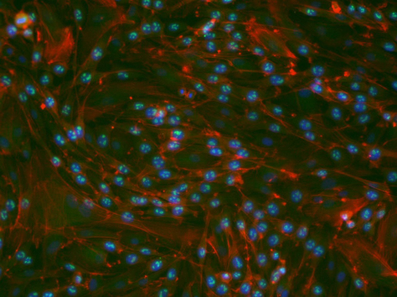
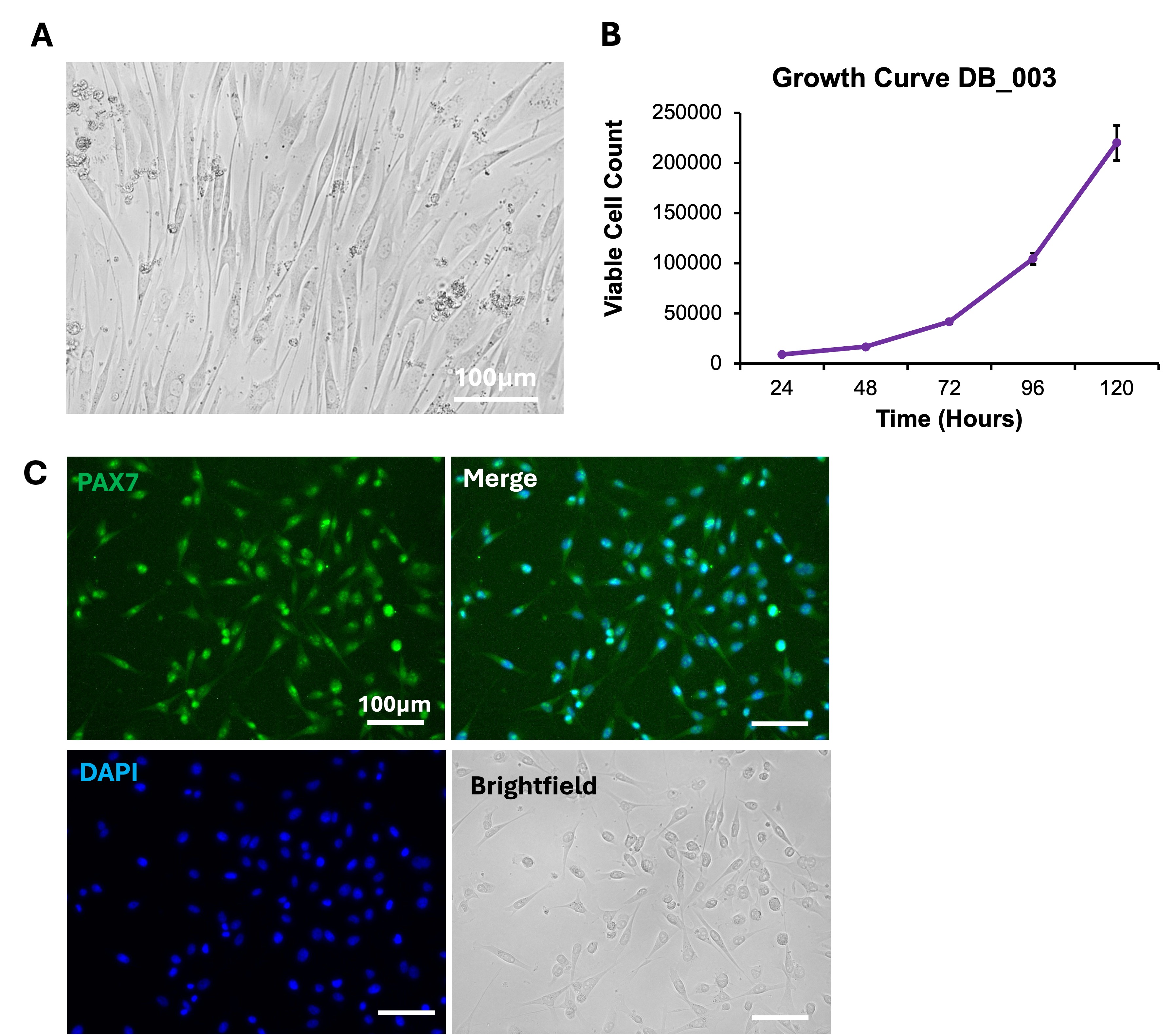
piMyoSats display a characteristic spindle-shaped morphology during adherent culture (Figure 1A) and demonstrate rapid proliferation over a 5-day growth period (Figure 1B). The cells retain skeletal muscle stem cell identity through expression of the canonical myogenic marker PAX7, confirmed by immunofluorescence staining (Figure 1C).
Myogenic Differentiation Characterisation
piMyoSats retain robust myogenic differentiation capacity and readily form multinucleated myotubes under differentiation conditions. Following a 7-day differentiation protocol, cells express MyosinHeavy Chain (MHC), a mature skeletal muscle marker, confirming successful myogenic differentiation and muscle fibre formation (Figure 2).
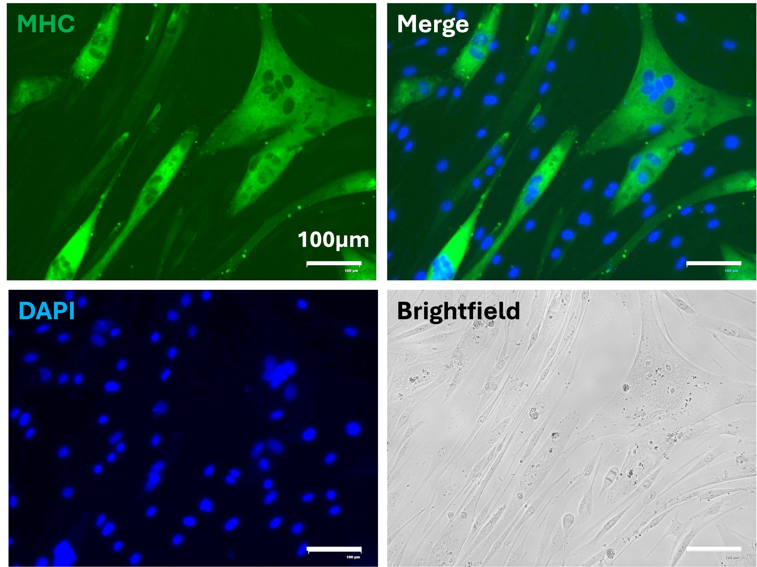
Representative immunofluorescence images of piMyoSats following 7 days of myogenic differentiation. Cells were stained for Myosin Heavy Chain (MHC) and DAPI nuclear counterstain. Robust MHC-positive staining confirms successful skeletal muscle differentiation and multinucleated myotube formation.
Compatible Processes
Product information
Cryopreserved cells, one vial containing 1x10⁶ cells

Species Specific Growth Factor Testing: QKine x Dragon
Species Specific Growth Factor Testing: QKine x Dragon