Porcine Induced Pluripotent Stem Cells
Porcine induced pluripotent stem cells (piPSCs) are reprogrammed from intramuscular fibroblasts and banked at P20+. These cells have been tested for sterility, mycoplasma, pluripotency marker expression, and tri-lineage differentiation through embryoid body assays. piPSCs can be differentiated using validated protocols into adipocytes and multinucleated skeletal muscle cells. piPSCs provide a consistent and reliable starting cell source for cultivated meat R&D, species-specific model development, and scalable production workflows.

Product Features
Versitile
Adapted for anchorage-dependent culture systems including plates, flasks, and microcarriers
Pluripotent
Able to differentiate into almost any cell type, including adipocytes, myocytes and myofibers.
Streamlined
Single starting cell for the creation of species-specific cell line models and product development
Cell Source and Reprogramming
This piPSC line was generated through non-integrative reprogramming of porcine intramuscular fibroblasts using the human transcription factors c-Myc, Klf4, Oct4, and Sox2. piPSCs are adapted to feeder-free culture using commercially available basement membrane extracts and demonstrate stable long-term proliferation while maintaining pluripotency.
Cell characterisation
piPSCs maintain typical pluripotent stem cell colony morphology and express key pluripotency markers including OCT4, NANOG, SOX2, and SSEA4 (Figure 1A–B). Cells exhibit rapid proliferation over a 5-day culture period, with an estimated doubling time of approximately 24 hours (Figure 1C).
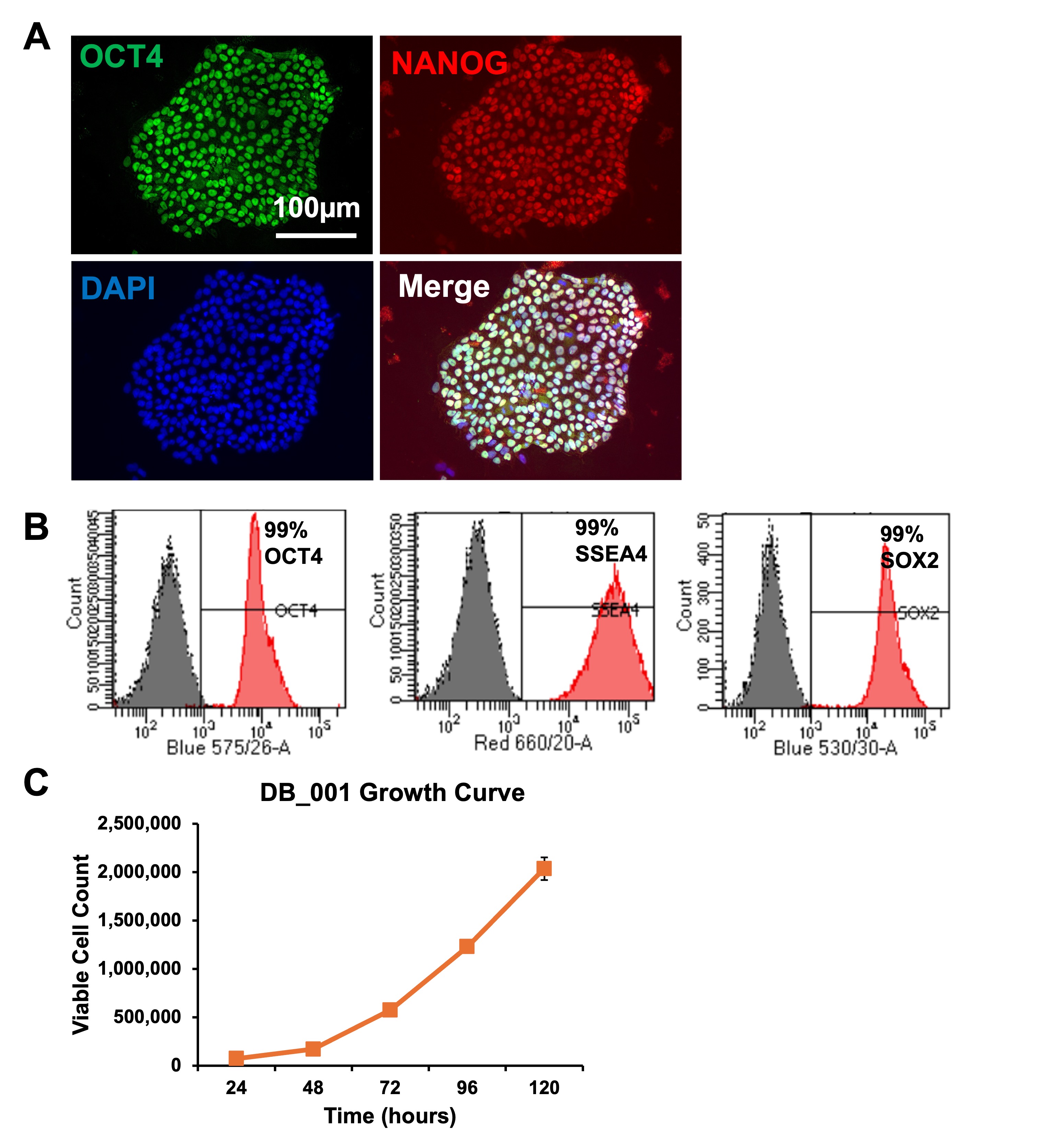
(A) Immunofluorescence staining showing expression of OCT4(green), NANOG (red), and DAPI (blue). (B) Flow cytometry analysis demonstrating expression of OCT4,SOX2, and SSEA4. (C) Growth curve indicating rapid proliferation over 5 days.
Differentiation Characterisation
piPSCs demonstrate robust tri-lineage differentiation capacity through embryoid body formation and directed differentiation protocols. Embryoid bodies express markers associated with ectoderm, mesoderm, and endoderm lineages. Directed differentiation protocols generates functional skeletal muscle cells expressing MyoD1 and MYH2, as well as adipocytes expressing PPARγ and CEBPA.
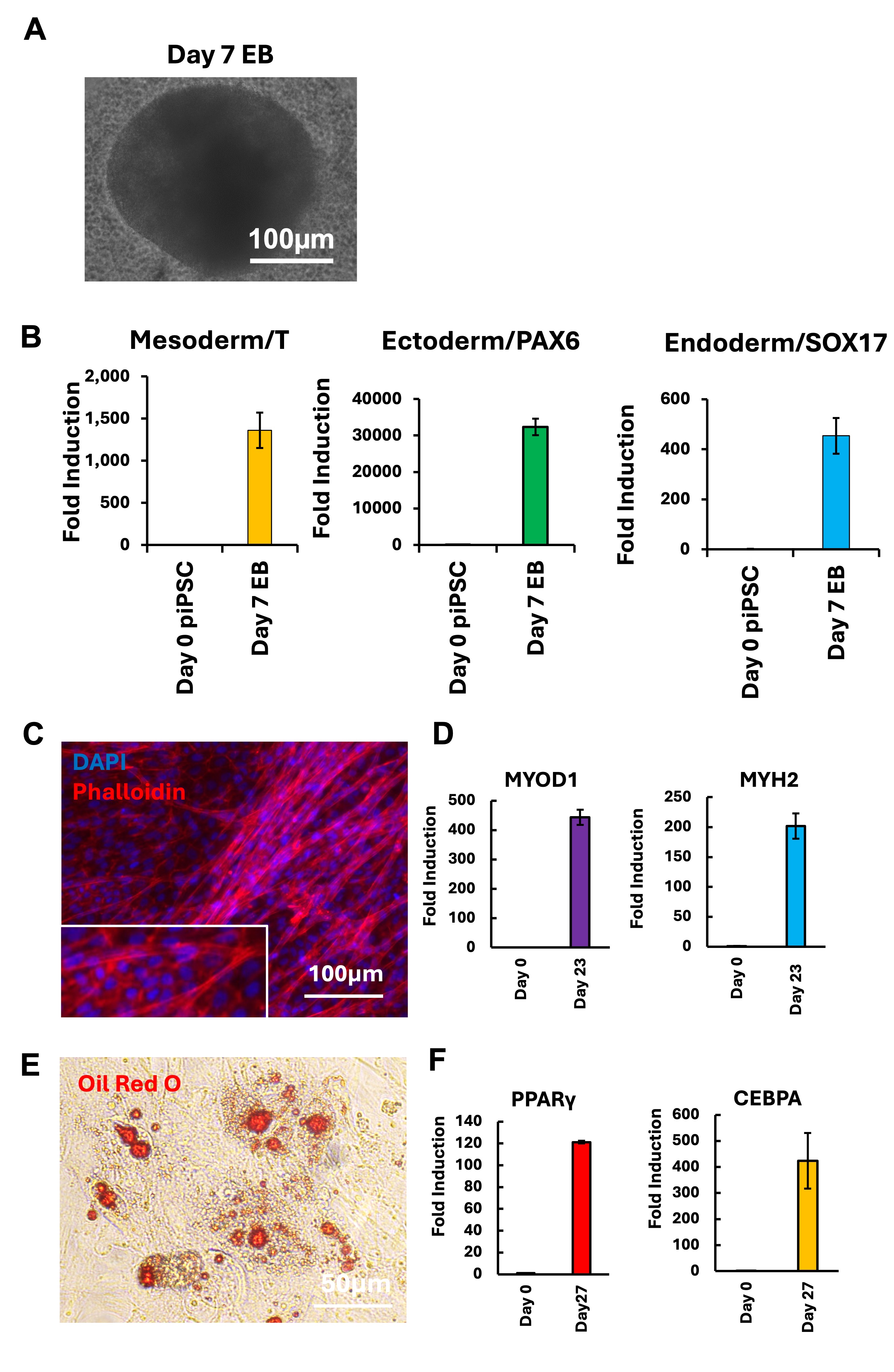
(A) Representative Day 7 embryoid body. (B) qPCR quantification of ectoderm, mesoderm, and endoderm markers. (C) Immunofluorescence microscopy of piPSC-derived skeletal muscle cells. (D) qPCR showing upregulation of MyoD1 and MYH2 muscle markers. (E) Oil Red O staining of piPSC-derived adipocytes. (F) qPCR showing upregulation of adipogenic markers PPARγ andCEBPA.
Microcarrier Suspension Adaptation
piPSCs readily attach to and expand on microcarrierswhile maintaining high viability under dynamic suspension conditions,supporting scale-up and bioprocess development applications

Compatible Processes
Product information
Cryopreserved cells, one vial containing appx. 2 x 10⁶ cells


Species Specific Growth Factor Testing: QKine x Dragon
Species Specific Growth Factor Testing: QKine x Dragon