Porcine spontaneously immortalised adipocyte derived stem cells
Porcine spontaneously immortalised adipose-derived stem cells (psiADSCs) are engineered for robust proliferation and efficient adipogenic differentiation. Adipose-derived stem cells (ADSCs) are the resident stem cell population found within adipose tissue. psiADSCs were generated using a proprietary non-integrative, non-GMO spontaneous immortalisation method, enabling long-term expansion while maintaining adipogenic functionality. These adherent cells exhibit rapid growth, with an average population doubling time of less than 17 hours and are optimised for scalable cultivated fat production, technology development and research applications.
Product Features
Non-GMO
Spontaneously Immortalised cell line using non integrative, non GMO techniques.
Immortalised
Functional immortalization ensures robust phenotype and performance over tens to hundreds of population doublings.
Efficient Adipogenic Differentiation
Maintains strong adipogenic lineage potential and readily differentiates into lipid-accumulating adipocytes.
Cell Source and Immortalisation
Primary porcine adipose-derived stem cells (ADSCs)were isolated from subcutaneous abdominal adipose tissue using enzymatic digestion methods. Cells were expanded on tissue culture-treated plastic for a minimum of two passages prior to immortalisation. Primary porcine ADSCs were subsequently subjected to a proprietary spontaneous immortalisation process designed to enable long-term proliferative capacity while preserving adipogenic functionality.
Cell characterisation
psiADSCs display characteristic adherent morphology during routine culture (Figure 1A) and demonstrate rapid proliferation over a 4-day growth period (Figure 1B).
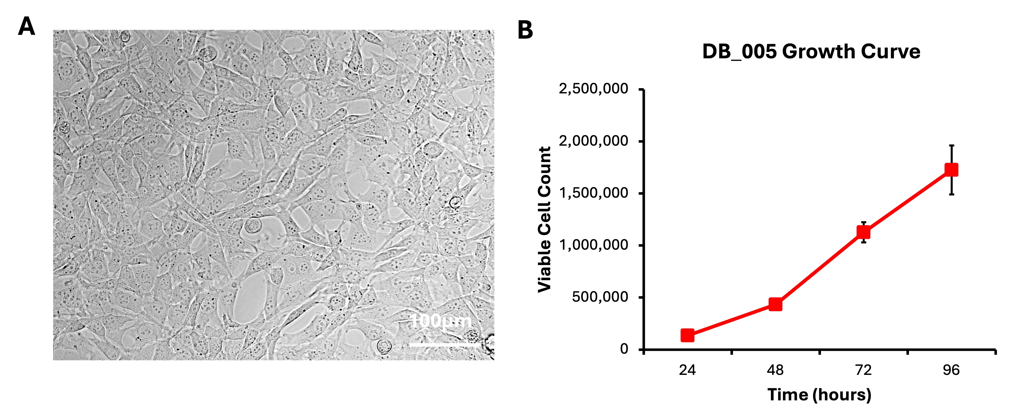
(A) Representative brightfield microscopy image showing psiADSC morphology.
(B) Growth curve of psiADSCs demonstrating robust proliferation over a 4-day period.
Adipogenic Differentiation Characterisation
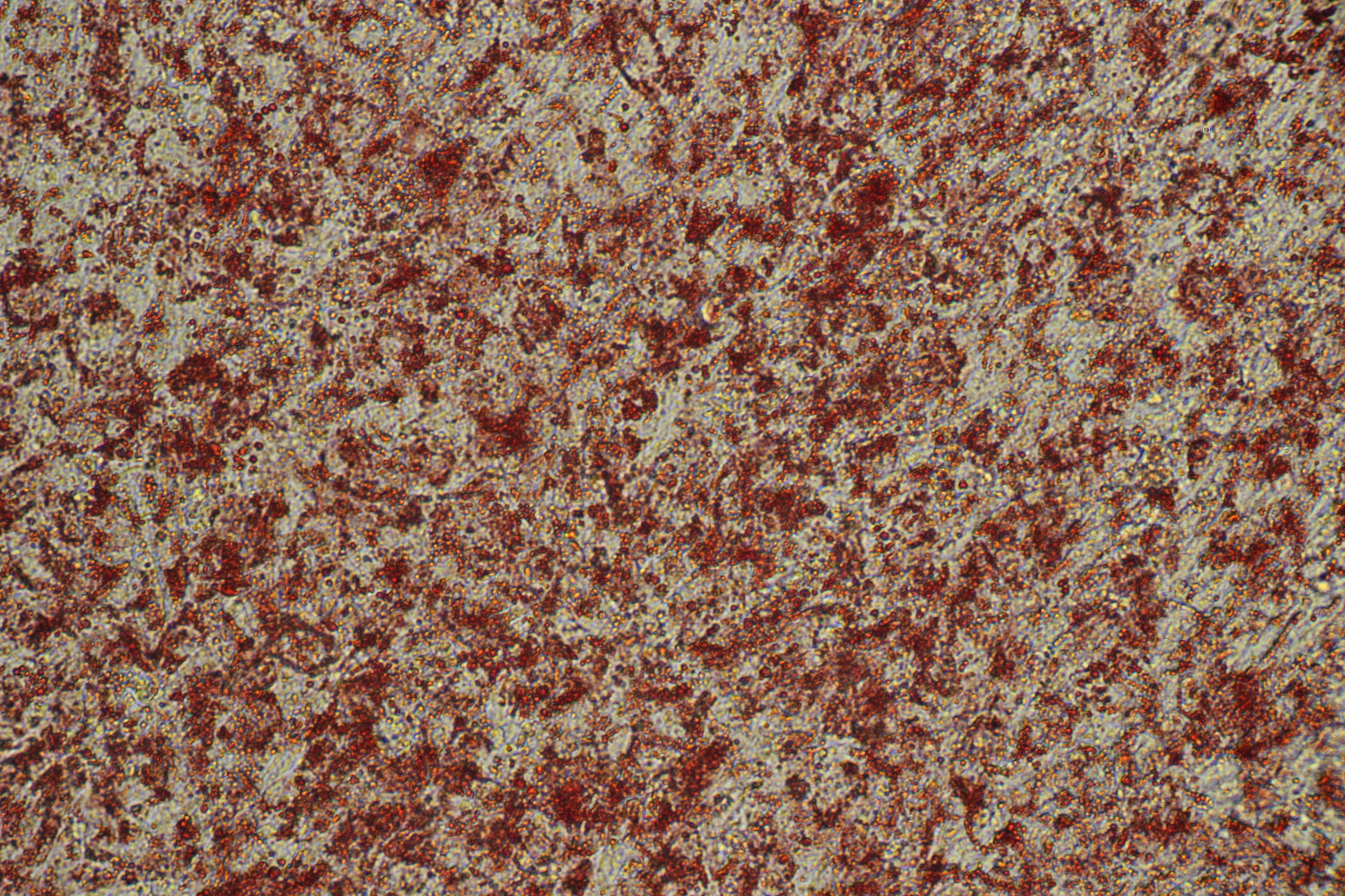
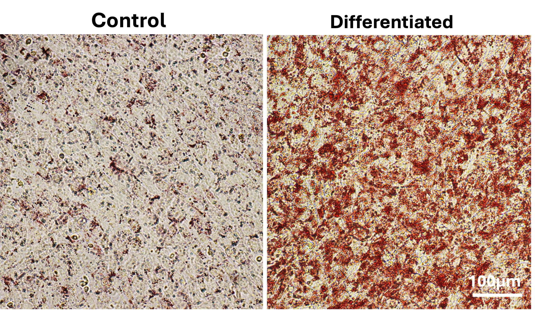
Importantly, psiADSCs retain strong adipogenic differentiation potential. Following a 10-day adipogenic differentiation protocol, cells develop intracellular lipid accumulation and differentiate into Oil Red O-positive adipocytes (Figure 2).
Compatible Processes
Product information
Cryopreserved cells, one vial containing appx. 1 x 10⁶ cells

Species Specific Growth Factor Testing: QKine x Dragon
Species Specific Growth Factor Testing: QKine x Dragon